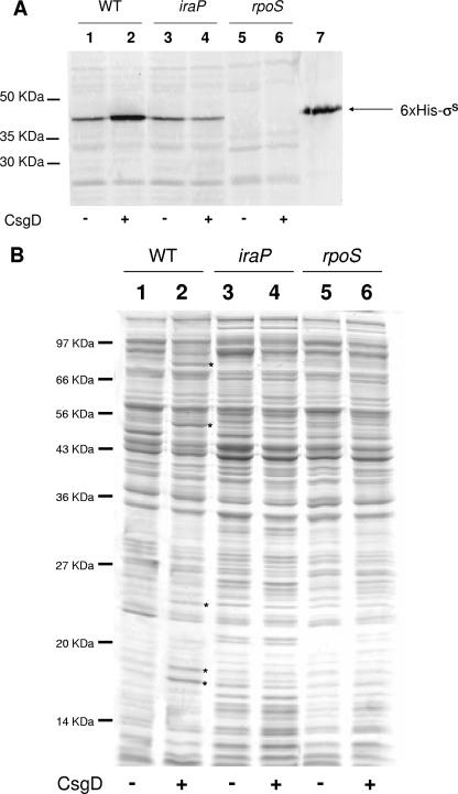
FIG. 6.
(A) Western blotting with anti-σS antibodies. Cell extracts were prepared from overnight cultures grown in M9Glu/sup at 30°C, and 20 μg of total proteins was loaded onto a SDS-polyacrylamide gel. Lane 1, MG1655/pT7-7; lane 2, MG1655/pT7CsgD; lane 3, LG03/pT7-7; lane 4, LG03/pT7CsgD; lane 5, EB1.3/pT7-7; lane 6, EB1.3/pT7CsgD; lane 7, purified His6-tagged σS. The positions of the 50-, 35-, and 30-kDa molecular mass markers are shown. (B) SDS-PAGE analysis of cytoplasmic proteins (20 μg of total proteins). Lane order is as described in panel A. Differently expressed proteins are indicated by an asterisk.

