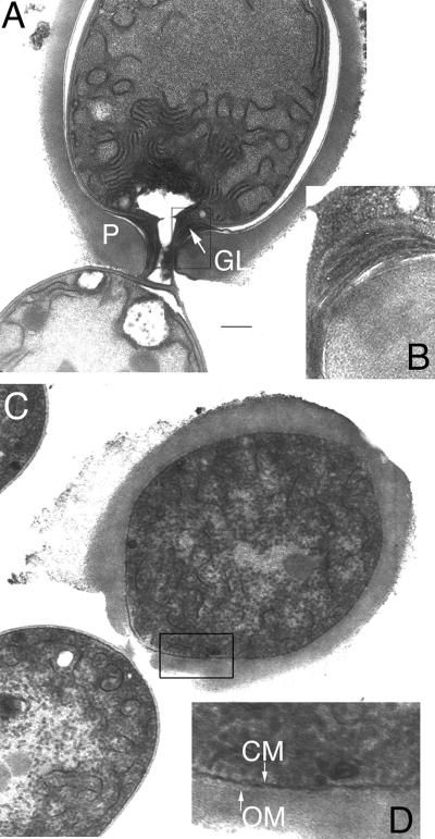
FIG. 3.
Ultrastructure of heterocysts of hgdD mutants: transmission electron micrographs of ultrathin sections of a connection of a heterocyst and a vegetative cell of the wild-type Anabaena sp. strain (A and B) and mutant DR181 (C and D). Magnifications of the cell wall and heterocyst envelope, indicated by squares in panels A and C, are shown for the wild-type strain and DR181 in panels B and D, respectively. In panel A, the white space surrounding the cell wall resulted from dehydration of the protoplast during sample preparation. Bar = 1 μm for panels A and C. GL, laminated layer; P, homogeneous layer; CM, cytoplasmic membrane; OM, outer membrane.

