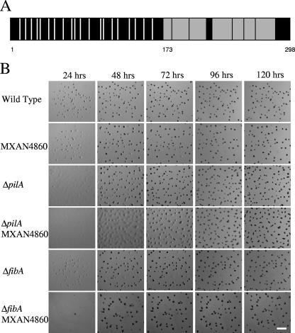
FIG. 2.
Primary structure analysis of MXAN4860 and developmental timing of MXAN4860 disruptions in wild-type and pilA strains. (A) Schematic of the MXAN4860 protein product. Vertical white bars indicate positions of 20 cysteine residues found in the N-terminal portion of the protein. Gray boxes indicate the tryptic peptides detected by mass spectrometry. The numbers indicate amino acid positions: 1, the start of the protein; 173, the start of the first peptide detected by mass spectrometry; 298, the end of the protein. (B) Developmental time course of fruiting body morphogenesis. Cells (5 × 10⁶) were spotted in 10 μl on TPM agar and photographed every 24 h. Strains with the MXAN4860 mutation develop normally unless it is coupled with a pilA mutation, in which case there is an approximately 24-h delay in development between the 48- and 72-h time points. Bar = 1 mm.

