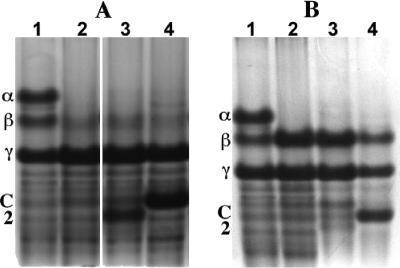
FIG. 1.
Acid-gel electrophoresis of SASP extracts from spores of various strains. Spores were prepared, purified, and extracted; the extracts were processed; aliquots were subjected to acid-gel electrophoresis; and the gels were stained, as described in the text. The extracts run in the various lanes were from spores of the following strains: (A) lane 1, PS533 (wild-type); lane 2, PS578 (α− β−); lane 3, PS4049 (α− β− Ssp2); and lane 4, PS1450 (α− β− SspC); (B) lane 1, PS533 (wild type); lane 2, PS260 (α−); lane 3, PS4051 (α− Ssp2); and lane 4, PS4052 (α− SspC). α, β, C, and 2, migration positions of SASP-α, SASP-β, SspC, and Ssp2, respectively; γ, migration position of the single γ-type SASP in B. subtilis spores. This last protein is very different from α/β-type SASP and plays no role in spore resistance properties (6, 13). The lanes in panel A are from the same gel, but several intervening lanes have been removed for clarity, and a white space was left to indicate where lanes were removed; the lanes in panel B are also from the same gel and were run adjacent to each other.

