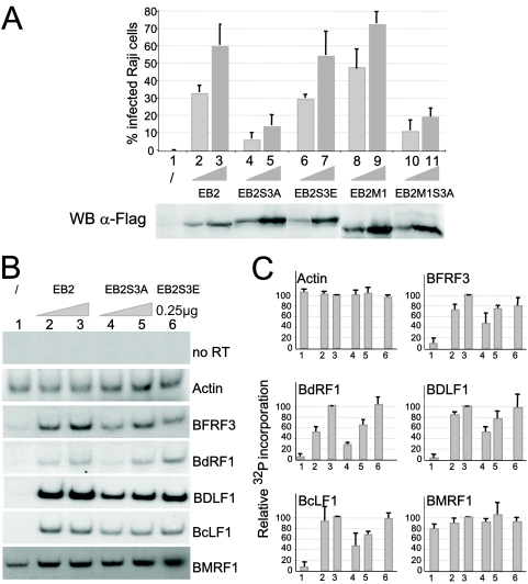
FIG. 7.
The EB2S3A mutant does not rescue infectious-virus production by 293BMLF1-KO cells and inefficiently exports several late EBV mRNAs. (A) 293BMLF1-KO cells were transfected with 1 μg of EB1 expression vector alone (lane 1) or together with 0.25 μg (lanes 2, 4, 6, 8, and 10) or with 0.5 μg (lanes 3, 5, 7, 9, and 11) of F.EB2, F.EB2S3A, F.EB2S3E, F.EB2M1, and F.EB2M1S3A expression vectors. Three days after transfection, Raji cells were infected with the filtered medium, and after 48 h, infected cells were quantified by FACS analysis (FACSCALIBUR). Two representative Raji cell infections are presented. All errors bars represent standard deviations. WB anti-FLAG, levels of expression of F.EB2 and F.EB2 mutated proteins in transfected 293BMLF1-KO cells, as evaluated by Western blotting with the M2 anti-FLAG MAb. (B) Cytoplasmic RNAs were extracted from 293BMLF1-KO cells transfected as described for panel A, except that RNA was extracted only from cells transfected with 0.25 μg of F.EB2S3E (lane 6). They were reverse transcribed using oligo(dT)18 as a primer and PCR amplified in the presence of 0.1 μCi [α-32P]dCTP with the specific primers shown in Table 1. The DNA fragments generated by PCR were separated on a 6% polyacrylamide gel and revealed by autoradiography. (C) The relative amounts of 32P incorporated in the PCR products presented in panel B were quantified using a PhosphorImager (Amersham). The results are presented as 32P incorporation relative to that obtained at the highest EB2 concentration, arbitrarily expressed as 100 (lanes 3). All errors bars represent standard deviations.

