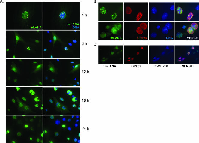
FIG. 2.
mLANA is a nuclear/cytoplasmic protein expressed during lytic replication. NIH 3T3 fibroblasts were infected with recombinant MHV68-73GFP at an MOI of 10 PFU/cell. (A) Cells were fixed at the indicated times postinfection and were stained with GFP-directed antiserum to visualize mLANA-GFP subcellular localization by fluorescence microscopy. (B and C) Cells were stained with antibodies to GFP or ORF59 18 h postinfection (B) or GFP, ORF59, and MHV68 antiserum 24 h postinfection (C). DNA was stained with DAPI (A) or Hoechst 33342 (B). The images in panel A at 4 h and in panel B were captured at ×100 magnification. All other images were captured at ×40 magnification.

