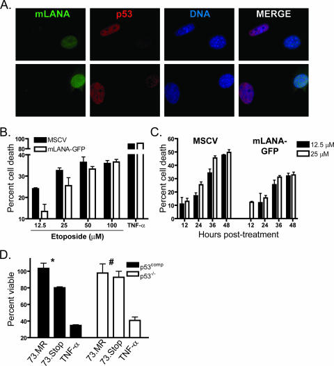
FIG. 6.
mLANA inhibits p53 induction and reduces etoposide-induced cell death. (A) mLANA-GFP-transduced NIH 3T3 fibroblasts were treated with etoposide (100 μM). Cells were fixed and stained for p53 4 h posttreatment. Two representative images are shown at ×100 magnification. (B and C) Empty vector (MSCV) or mLANA-GFP-transduced NIH 3T3 fibroblasts were treated with etoposide at the indicated concentrations. Cell viability was analyzed 28 h posttreatment (B) or during a time course (C). Data are presented as the percentages of cell death with treatment relative to that for mock-treated controls. Results are the means from triplicate samples. Error bars represent standard deviations. (D) p53comp or p53−/− MEFs were infected with 73.Stop or 73.MR virus at an MOI of 2 PFU/cell in the presence of 20 ng/ml cidofovir. Cell viability was determined 12 h postinfection. Data represent the percentages of cell death relative to that for mock-infected samples. Results are the means from triplicate samples. Error bars represent standard errors of the means. Statistical analyses of differences for 73.MR- and 73.Stop-infected cells were performed using a two-tailed paired Student's t test (*, P = 0.05; #, P = 0.66). (B and D) Tumor necrosis factor alpha (TNF-α) samples were treated with 25 ng/ml TNF-α and 10 μg/ml cycloheximide as a positive control for cell death induction.

