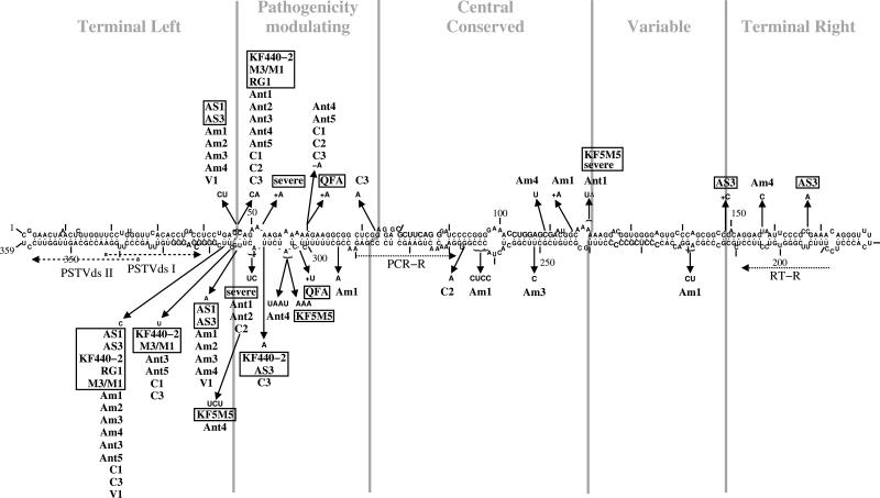
FIG. 5.
Localization of mutations in PSTVds from infected weeds. The secondary structure is based on consensus base-pairing probability of all PSTVd strains used for infection and resulting mutants found in the present study. The sequence of the PSTVd strain “intermediate” (9) is given. Nucleotide positions that differ in the strains and mutants from the “intermediate” sequence are marked by arrows. The names of PSTVd strains used for infecting weeds are boxed. Individual secondary structures of mutants and infecting, parental strains are available (see Fig. S1 at http://www.biophys.uni-duesseldorf.de/Matousek_JVI_2007/). Am1 to Am4, mutants isolated from Amaranthus retroflexus (redroot amaranth); Ant1 to Ant5, Anthemis arvensis (corn chamomile); C1 to C3, Chamomilla recutita (German chamomile); V1, Veronica arvensis (common speedwell). In gray are marked the domains in viroids (16). Primers used for cloning of mutants (PSTVdsI and PSTVdsII) and for quantification by real-time PCR (PCR-R and RT-R) are marked by dotted arrows.

