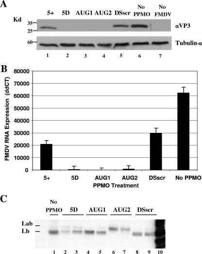
FIG. 4.
Effect of PPMO treatment on viral protein and RNA expression. (A) Western blot prepared from cell lysates obtained from BHK-21 cells treated with 2.5 μM of the indicated PPMO. Cell monolayers were PPMO-treated for 3 h and infected with FMDV A24Cru at a MOI of 5 PFU/cell for 1 h, followed by a further 4 h of incubation in a PPMO mixture before cell lysates were prepared (see Materials and Methods). Ten micrograms of total protein was loaded per lane, and the blot was probed with a polyclonal antibody for FMDV VP3 (upper panel) or a monoclonal antibody against tubulin-α (lower panel). (B) Quantitative real-time PCR measurement of FMDV RNA isolated from cell lysates corresponding to those in the above experiment. Values are reported as ΔΔCT, normalized to that of endogenous 18S rRNA and calibrated against mock-treated uninfected control cell lysates. The y axis indicates ΔΔCT (log2 relative units). Each error bar refers to the standard deviation for the respective mean data point (n = 3). Similar results were obtained in at least two independent experiments. (C) FMDV translation products produced in BHK-21 S10 lysates in the presence or absence of PPMO. Translation reactions were programmed with full-length A24Cru RNA in the presence of [35S]-methionine and immunoprecipitated with a polyclonal antibody for Leader protein. PPMOs were included in the translation reactions at concentrations of 0 μM (lane 1), 0.25 μM (lanes 3, 4, 6, and 8), and 2.5 μM (lanes 2, 5, 7, and 9). Lane 10 contains non-PPMO-treated and nonimmunoprecipitated viral RNA translation products. The products were examined by SDS-PAGE on a 12% gel.

