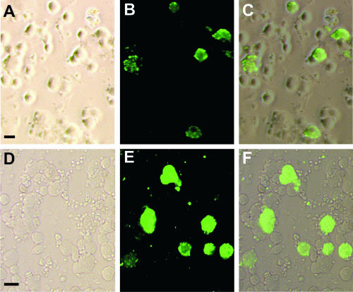
FIG. 1.
DRG neurons from wild-type mice infected by RV. (A to C) Neurons cultured for 16 h and subsequently infected for 20 h; (D to E) DRG neurons infected in vivo 6 days earlier and subsequently cultured for 4 h. Pictures were taken from areas containing many infected neurons to illustrate their appearance rather than to provide a representative picture of the percentage of infected cells. (A) Phase-contrast image; (D) bright-field image. (B and E) Immunoreactivity for RV phosphoprotein; (C and F) overlay of both pictures. Only large cells are infected in vitro or in vivo. Bars, 50 μm.

