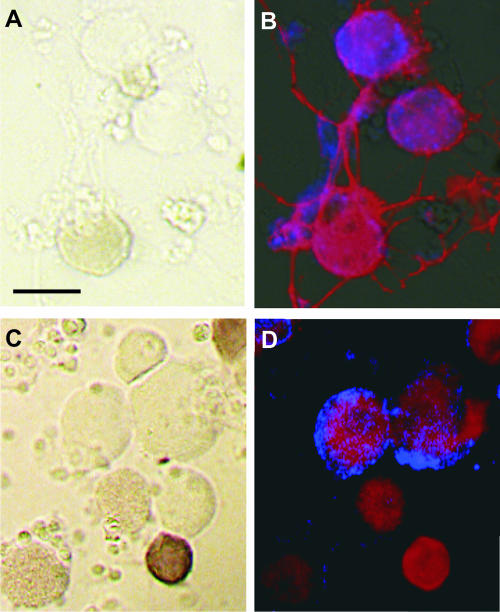
FIG. 2.
DRG neurons cultured for 16 h and subsequently infected with RV (A and B) for 12 h or exposed to RV (C and D), each for 30 min, before further processing. (A and C) Bright-field images; (B and D) fluorescent images. (A and C) Dark brown cells are neurons showing capsaicin-induced cobalt uptake. (B and D) Immunoreactivity for p75NTR (red, extending through the whole soma) and RV phosphoprotein (blue). Bars, 50 μm.

