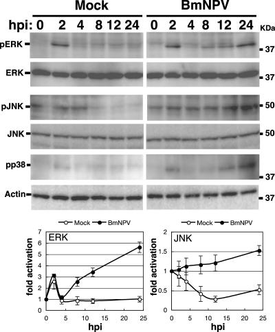
FIG. 5.
Activation of ERK and JNK by BmNPV in BmN cells. BmN cells were mock infected or infected with BmNPV at an MOI of 5. Activation of ERK and JNK at 0, 2, 4, 8, 12, and 24 hpi was assessed by Western blotting using phospho-ERK (pERK)- and phospho-JNK (pJNK)-specific antibodies, respectively. Total levels of ERK and JNK were examined by anti-ERK and anti-JNK antibodies, respectively. Western blots using anti-phospho-p38 (pp38) and anti-actin were also shown. MAPK activation plots show means ± SE of quantitative data from four to six independent experiments.

