
FIG. 5.
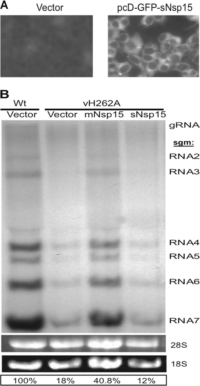
RNA accumulation in Nsp15-trans-complemented DBT cells infected with vH262A. (A) Detection of sNsp15 expression. DBT cells were transfected either with vector only or with pcD-GFP-sNsp15 and were subjected to GFP detection by fluorescent microscopy of cells at 30 h posttransfection. (B) DBT cells were first transfected either with vector only or with a plasmid driving the expression of Nsp15 derived from MHV-A59 or SARS-CoV (pcD-mNsp15 or pcD-sNsp15) and then infected with vH262A or WT virus 30 h later. At 6 hpi, viral RNAs were metabolically labeled as described in Materials and Methods. The labeled viral RNAs were resolved by formaldehyde agarose gel electrophoresis and visualized by autoradiography. The identities of the MHV RNAs are given on the right.