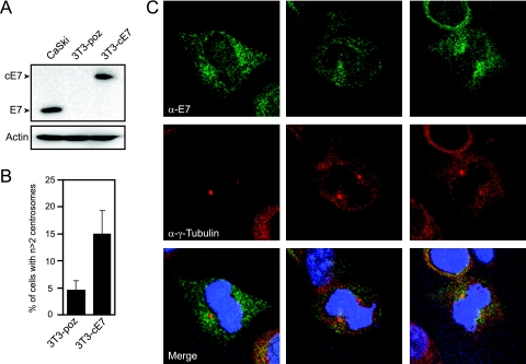
FIG. 1.
HPV16 E7 can concentrate around mitotic spindle poles. (A) Western blot analysis of E7 expression levels in stable NIH 3T3, 3T3-poz (control), and 3T3-cE7 cells compared to HPV-positive CaSki cervical cancer cells. Actin is shown as a loading control. (B) Percentage of 3T3-cE7 cells with more than two centrosomes compared to control 3T3-poz cells. Bar graphs indicate averages of four experiments in which >150 cells were counted per experiment. Error bars indicate the standard errors between experiments. (C) Representative images of mitotic NIH 3T3 cells with stable expression of C-terminally HA- and FLAG-tagged HPV16 E7. Coimmunofluorescence was performed using an E7-specific antibody (α-E7) and a γ-tubulin antibody (α-γ-Tubulin) as a centrosomal marker. Nuclei were visualized with Hoechst 33258 DNA dye.

