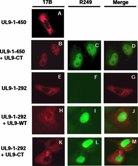
FIG. 5.
Confirmation of the N- and C-terminal interaction by immunofluorescence microscopy analysis. Vero cells were transfected with UL9-1-450 (A), UL9-1-450 plus UL9-CT (B, C, and D), UL9-1-292 (E, F, and G), UL9-1-292 plus UL9-WT (H, I, and J), or UL9-1-292 plus UL9-CT (K, L, and M). Panels A, B, E, H, and K were stained with the 17B antibody (recognizes the N-terminal 33 aa of UL9). Panels C, F, I, and L were stained with the R249 antibody (directed against the C terminus of UL9). Panels A and E show that UL9-1-450 and UL9-1-292 stayed in the cytoplasm. Panels C, I, and L show that UL9-WT and UL9-CT went to the nucleus. Panel B shows that the N-terminal fragment UL9-1-450 went to the nucleus along with UL9-CT. Panels H and K show that UL9-1-292 stayed in the cytoplasm, even in the presence of UL9-WT or UL9-CT.

