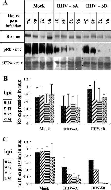
FIG. 4.
Rb hypophosphorylation in HHV-6A- and HHV-6B-infected SupT1 cells. (A) Western blots of mock-infected and virus-infected nuclear lysates were reacted with antibodies to Rb and pRb proteins. (B and C) Graphic representation of the results relative to the highest level of Rb or pRb protein found in the samples. The intensities of Rb and pRb were corrected for loading variations by the intensity of eIF2α. The measurements for the Rb were based on results from two experiments. The comparison of the pRb in the mock infection versus that of HHV-6B (Z29) infection was based on results from a single experiment. pRb in the cytoplasmic fractions of mock-infected and virus-infected cells was not detected (data not shown). The quantitations of pRb in the mock and HHV-6A (U1102) infections were based on results from three separate experiments. nuc, nuclear.

