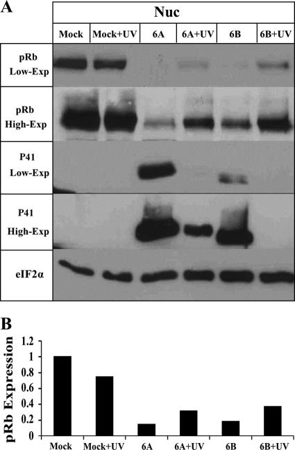
FIG. 6.
Dephosphorylation of Rb in cells infected with inactivated virus. (A) The viruses were irradiated by UV or were untreated, and Western blots from mock-infected and virus-infected cell lysates were reacted with antibodies to the pRb and P41 proteins (9A5 MAb). Two exposures of the blots (low-exp and high-exp) are shown for the pRb and the P41 proteins. (B) Graphic representation of the results relative to the highest pRb levels (mock-infected cells). The intensities of pRb were corrected for loading variations by the intensity of eIF2α. nuc, nuclear.

