FIG. 3.
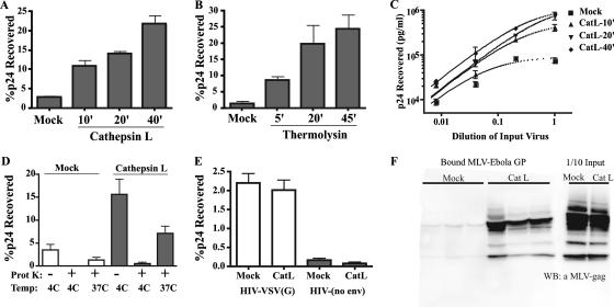
Proteolytic cleavage of Ebola virus GP enhances binding of virions to cells. HIV-Ebola virus GP pseudovirions were not treated (mock) or were treated with CatL (A) or thermolysin (B) at 37°C. Proteolytic reactions were terminated at the indicated times, and virions were added to VeroE6 cells chilled to 4°C. Virions were bound to cells by centrifugation. Unbound virus was removed by washing cells in medium at 4°C. Cell lysates were analyzed by p24 antigen ELISA. Results are the percentages of input p24 recovered after binding and are the means and standard deviations for samples run in triplicate. (C) HIV-Ebola virus GP virions were mock treated or treated with CatL for the indicated times, serially diluted fivefold, and bound to VeroE6 cells as described for panel A. Results are concentrations of p24 recovered in cell lysates and are the means and standard deviations for samples run in triplicate. (D) As a control for virus internalization during binding, mock- and CatL-treated virions were bound to cells at 4°C, washed, and lysed immediately or treated with 50 μg/ml proteinase K prior to lysis. Alternatively, cells with bound virions were incubated at 37°C for 30 min followed by treatment with proteinase K and cell lysis. Results are presented as in panel A. Similar results were obtained with 293T cells (data not shown). (E) HIV-VSV(G) pseudovirions and nonenveloped HIV core particles were mock treated or treated with CatL for 30 min. Virus preparations were bound to cells, washed, and lysed. The amount of cell-associated virus was quantified by p24 antigen ELISA. Results are percentages of input p24 recovered after binding and are the means and standard deviations for samples run in triplicate. (F) MLV-Ebola virus GP pseudovirions were mock treated or treated with CatL for 30 min. Virions were bound to cells, washed, and lysed. Samples were subjected to 4 to 15% sodium dodecyl sulfate-polyacrylamide gel and immunoblotted using polyclonal MLV Gag antisera. One-tenth of the virus input added to cells was directly analyzed by Western blotting to compare the level of virions present after proteolysis. Bound mock- and CatL-treated MLV-Ebola virus GP represent independent samples run in triplicate.

