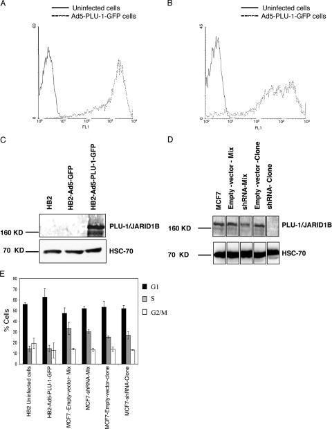
FIG. 1.
Regulation of PLU-1/JARID1B expression in human mammary epithelial cells using a recombinant adenovirus and shRNA. (A and B) Uninfected or Ad5-PLU-1-GFP-infected HB2 cells were incubated for 24 (A) or 72 (B) hours and analyzed by flow cytometry. The y axis depicts cell number, and the x axis depicts log relative fluorescence intensity (FL1) for GFP expression. The graph of HB2 cells infected with the Ad5-GFP control vector is not shown because the reading was extremely bright and partially off the scale. (C) Whole-cell extracts prepared from HB2 cells infected for 24 h with Ad5-GFP or Ad5-PLU-1-GFP or uninfected were separated by SDS-PAGE and immunoblotted using α-PLU-1-C antiserum (upper panel) or anti-HSC70 antibody (lower panel) to check for protein load. (D) Western blot analysis of whole-cell protein lysate of shRNA-transfected MCF7 cell lines or the corresponding empty vector transfectants. The upper panel shows the immunoblot analysis using α-PLU-1-C antiserum, while the lower panel shows the immunoblot analysis using anti-HSC70 antibody. (E). Changing the level of expression of the PLU-1/JARID1B gene does not alter the cell cycle profile of HB2 and MCF7 cells. HB2 cells that were noninfected or infected for 48 h with Ad5-PLU-1-GFP and MCF7 cells stably transfected with an empty vector (MCF7-empty-vector-clone and MCF7-empty-vector-mix) or with a PLU-1-specific shRNA expression vector (MCF7-shRNA-clone and MCF7-shRNA-mix) were subjected to cell cycle analysis (BrdU incorporation and Hoechst staining) as described in Materials and Methods. Bars represent the means ± standard errors of the means of three independent experiments.

