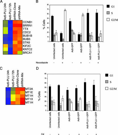
FIG. 8.
Overexpression of PLU-1/JARID1B attenuates the G2/M block induced by treatment with either of the microtubule-destabilizing agents nocodazole or cadmium. (A and C) Color-coded TreeView diagrams depicting genes significantly downregulated following the upregulation of PLU-1/JARID1B expression. For details, see the legend for Fig. 2A. (B and D) HB2 cells were infected at an MOI of 500 with no adenovirus (uninfected cells), with the adenovirus GFP vector (Ad5-GFP), or with the adenovirus recombinant vector expressing a GFP-tagged PLU-1/JARID1B gene (Ad5-PLU-1-GFP). After 24 h of infection, cells were treated (+) or not treated (−) with 0.4 μg/ml nocodazole (B) or with 20 μM CdCl2 (D) for a further 24 h. At the end of the incubation, cells were pulse labeled (30 min) with 10 μM BrdU, then harvested, fixed in 1% paraformaldehyde, and stained for DNA content (Hoechst/Triton X-100) and BrdU incorporation (Alexa Fluor 697 intensity) analysis. In uninfected cells, the Hoechst versus BrdU plots were used to assess percentages of cells in G1, S, and G2. In infected cells, profiles were first gated on GFP-positive events before the cell cycle phases were derived. Bars represent the means ± standard errors of the means of three independent experiments.

