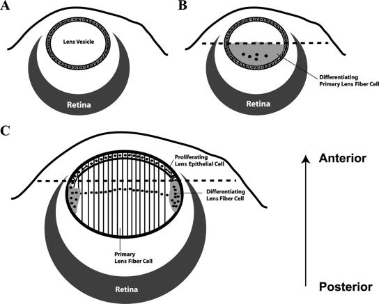
FIG. 1.
Diagrams showing different developmental stages of the ocular lens. (A) Lens vesicle. (B) Formation of primary lens fiber cells. (C) Formation of secondary lens fiber cells. Dashed lines in panels B and C represent the boundary of differentiation, epithelial cells anterior to which are not induced to differentiate. The arrow (posterior to anterior) represents the orientation of all lens images throughout this paper.

