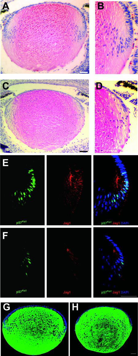
FIG. 8.
Formation of secondary lens fiber cells is decreased in Notch signaling mutants. (A) Microphotograph of a hematoxylin-and-eosin-stained E17.5 control lens section. (B) Higher magnification of the transition zone shown in panel A. (C) Microphotograph of an hematoxylin-and-eosin-stained E17.5 Rbp-J mutant lens section. (D) Higher magnification of the transition zone shown in panel C. (E and F) Immunofluorescent Jag1 and p57Kip2 staining in section of control (E) and Rbp-J mutant (F) lenses. (G and H) Immunofluorescent β-crystallin staining in sections of control (G) and Rbp-J mutant (H) lenses. Sections in panels E to H were counterstained for DNA with DAPI. Scale bars, 50 μm.

