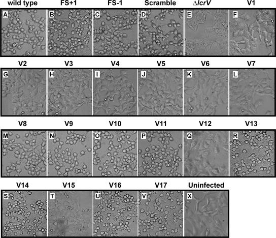
FIG. 4.
Infection of HeLa cells by different strains of Y. pseudotuberculosis. At 2 h of infection, the effect of the bacteria on HeLa cells was recorded by phase-contrast microscopy. The experiment was repeated at least three times. Note the extensive rounding up of the YopE-dependent, cytotoxically affected HeLa cells (A to D, M to P, R, S, U, and V). Shown are phase-contrast images. The strain designations are identical to those used in Fig. 1. Panel X is an uninfected HeLa cell monolayer control.

