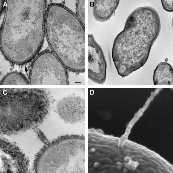
FIG. 4.
Ultrastructural evidence of extracellular polysaccharide capsule and conjugative pilus-like structures. (A and B) Transmission electron micrographs of G. bethesdensis NIH1.1 prepared with (A) and without (B) ruthenium red for preserving and contrasting extracellular polysaccharides. The arrow in panel A points out the capsular material on the surface. (C and D) Transmission (C) and scanning (D) electron micrographs of structures resembling conjugative pili. Bars, 100 nm.

