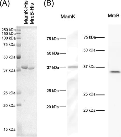
FIG. 1.
(A) Sodium dodecyl sulfate-polyacrylamide gel electrophoresis profile of the purified C-terminal His-tagged MamK and MreB from E. coli C41(DE3). The protein bands were stained with Coomassie brilliant blue G-250. (B) Immunoblotting analyses of M. magnetotacticum cell extract with the anti-MamK and the anti-MreB antibodies. The antibodies generated against MamK and MreB showed monospecificities as single positive bands with apparent molecular masses of 38 kDa and 36 kDa, respectively. The apparent molecular masses of the positive bands corresponded to the deduced molecular masses from the mamK and mreB genes. Proteins (10 μg/lane) extracted from M. magnetotacticum cells were loaded on each lane. The molecular masses of the standards (Precision Plus protein standards; Bio-Rad) are indicated on the left sides of the lanes.

