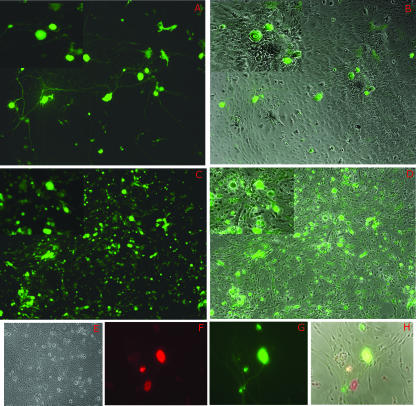
FIG. 2.
d109 transgene expression in fetal TG cultures. Primary cultures of embryonic TG were infected with either d109 (A, B, and F to H) or d106 (C and D) and photographed 24 h postinfection. Duplicate exposures of the same field showing either phase-contrast results together with GFP expression or GFP expression alone are shown. In order to provide greater detail, magnified insets of all fields are shown in the upper left corner of each exposure. (F, G, and H) Triplicate exposures of the same field showing either anti-HuC/D (neuron-specific antibody) expression alone (red in panel F), GFP expression alone (G), or phase-contrast exposure together with anti-HuC/D and GFP expression (H). (E) Mock-infected embryonic TG.

