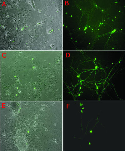
FIG. 3.
Persistence of d109 in neuron cultures. Cultures of embryonic TG were infected with d109 and photographed over time. Shown are duplicate exposures of d109 infection depicting either phase-contrast results and GFP expression or GFP expression alone. d109 is shown at day 1 postinfection (A and B), at day 7 postinfection (C and D), and at day 38 postinfection (E and F).

