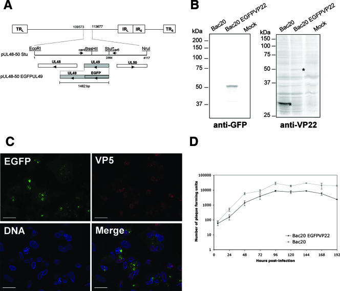
FIG. 1.
Generation and characterization of recombinant MDV expressing EGFPVP22. (A) Schematic representation of the MDV genome. The 4,117-bp EcoRI-NruI fragment (positions 109573 to 113677 in the Md5 sequence, accession no. AF243438), containing an StuI restriction site inserted immediately upstream of the UL49 start codon, was cloned into pGEM-Te to yield plasmid pUL48-50 Stu. This plasmid contains UL49 and its flanking sequences derived from the RB1B genome. Arrowheads indicate the direction of gene transcription. The construct pUL48-50 EGFPUL49 containing the EGFP coding region fused to the 5′ end of the MDV UL49 open reading frame in the same plasmid backbone is also shown. (B) Analysis of EGFPVP22 protein expression by Western blotting. CESCs infected with the Bac20 EGFPVP22 or the parental Bac20 virus were trypsinized 5 days postinfection, pelleted by centrifugation, and directly lysed in 2× Laemmli buffer. Sonicated crude cell lysates were separated by sodium dodecyl sulfate-polyacrylamide gel electrophoresis on a 10% polyacrylamide gel and transferred to nitrocellulose. The blots were incubated with the rabbit anti-GFP antibody or the L13a anti-VP22 MAb. Mock corresponds to noninfected cells. The molecular mass marker positions (molecular mass are in kilodaltons) are indicated on the left. The molecular mass of the EGFPVP22 protein was estimated on the gel at 50 kDa. (C) Fluorescence analysis on CESCs infected with the Bac20 EGFPVP22 virus. Cells were infected on coverslips and fixed at 5 days postinfection. EGFPVP22 was visualized via EGFP fluorescence (green), the VP5 major capsid protein was stained with the F19 MAb (red), and the nuclei were stained with Hoechst 33342 dye (blue). Cells were examined under a Zeiss microscope with the ApoTome system. EGFPVP22 was found in both the cytoplasmic and nuclear compartments. Bars, 20 μm. (D) Growth of Bac20 EGFPVP22 compared to that of the Bac20 parental virus. Approximately 5 × 10⁵ CESCs were infected with 100 PFU of the respective MDV. At 6, 24, 48, 72, 96, 120, 144, 168, and 192 h postinfection, cells were trypsinized and titers were determined on fresh CESCs (two wells per dilution). Virus plaques were counted at 5 days postinfection under a light microscope. The curves represent the mean value of two independent experiments; error bars indicate the standard deviation of the mean.

