FIG. 4.
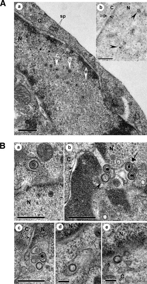
Nucleocapsids and egress from the nucleus. (A) The three types of capsids (A, B, and C) were detected in the nucleus. In this picture, the C nucleocapsids range from 94 to 102 nm in diameter. Small round particles, termed small particles (sp), frequently reported to be associated with herpesvirus nuclear capsids are indicated by arrows. The inset (part b) shows nuclear capsids exhibiting strong labeling with an anti-VP5 MAb (black arrowheads). (B) Egress from the nucleus. In part a, one single enveloped B nucleocapsid was observed in the perinuclear space as the vacuole membrane seemed to be a continuation of the outer nuclear membrane. This primary enveloped virion had a diameter of approximately 150 nm. Part b shows the accumulation of four enveloped MDV particles in the perinuclear cisterna, including a capsidless particle (black arrowhead) and three particles with an electron-dense core (C capsids). For one of these virions, the primary envelope is in continuity with the inner nuclear envelope (black arrow). The enveloped particles with capsids ranged from 131 to 157 nm in diameter. In part c, an additional image of such perinuclear accumulation of particles is shown. In parts d and e, capsidless particles in a vacuole in proximity to the nucleus and fused to the outer membrane of the nuclear envelope are shown. N, nucleus; C, cytoplasm. The inner membrane (arrow 1) and outer membrane (arrow 2) of the nuclear envelope are indicated.

