FIG. 1.
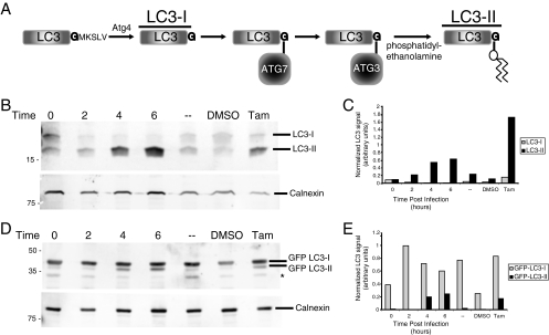
Modification of LC3 protein during poliovirus infection. (A) Schematic representation of LC3 processing as it occurs during autophagy activation. LC3-I is the unmodified, cleaved form of LC3, while LC3-II is the lipid-conjugated species. (B and C) Human 293T cells were either infected with poliovirus at a multiplicity of infection of 50 PFU per cell and harvested at 0, 2, 4, or 6 h after infection, mock infected (-), treated with 10 μM tamoxifen, or treated with control solvent (dimethyl sulfoxide [DMSO]) for 48 h prior to harvesting. (B) Cell extracts were subjected to PAGE through a 13.5% gel, and separated proteins were immunoblotted using a rabbit polyclonal anti-LC3 antibody and subsequently detected with goat anti-rabbit alkaline phosphatase and ECF reagent (Amersham Biosciences). The same immunoblots were reprobed with a mouse monoclonal anticalnexin antibody. (C) The amount of signal for both endogenous LC3-I and endogenous LC3-II from panel B is presented graphically. (D) Cells were transfected with a plasmid that expressed GFP-LC3 for 48 h before infection. Cell extracts were subjected to PAGE in a 10% polyacrylamide-SDS gel, and separated proteins were immunoblotted for LC3 and calnexin as for panel B. The asterisk signifies an unidentified cross-reacting band. (E) The amount of signal for GFP-LC3-I and GFP-LC3-II from panel D is presented graphically. The gels are representative of multiple experiments.

