FIG. 1.
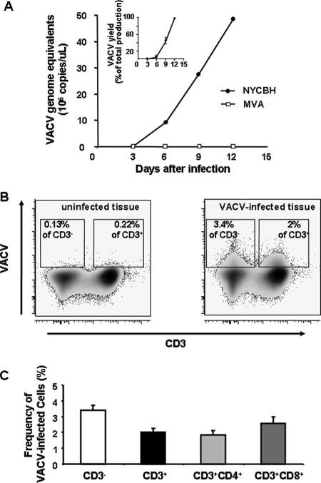
Replication of VACV in human lymphoid tissue ex vivo. Tissue blocks (27 from each human tonsil) were infected with either NYCBH or MVA. Each of the 27 tissue blocks from a single donor was inoculated with 5 μl of viral stock containing approximately 1 × 10⁶ genome equivalents. The culture medium was changed every 3 days, and VACV replication was monitored in these medium samples by means of real-time PCR (A) or flow cytometry (B and C). A. Typical replication kinetics of NYCBH and MVA are presented. The inset represents the average kinetics of replication of NYCBH (n = 13); results are shown as means ± SEM. Each point represents the measurement of medium pooled from three wells, each of which contained nine tissue blocks. To merge the data from different experiments, we normalized the amount of VACV DNA for each experiment as a percentage of the maximum production of VACV over the 12-day period. B. Expression of VACV antigen by lymphocytes as detected with flow cytometry. Density plots of lymphocytes, isolated from VACV-infected (right panel) and matched uninfected tissue (left panel), indicate preferential staining of CD3− cells for cell-associated vaccinia virus antigen on day 12 postinfection. One representative experiment (out of 11) is shown. C. Frequencies of VACV-infected CD3−, CD3+, CD3/CD4+, and CD3/CD8+ cells detected at day 12 postinfection. Cells were stained at day 12 for CD3, CD4, CD8, and cell-associated vaccinia virus antigens. Data are presented as means ± SEM (error bars) of VACV-infected cells from 11 donors.

