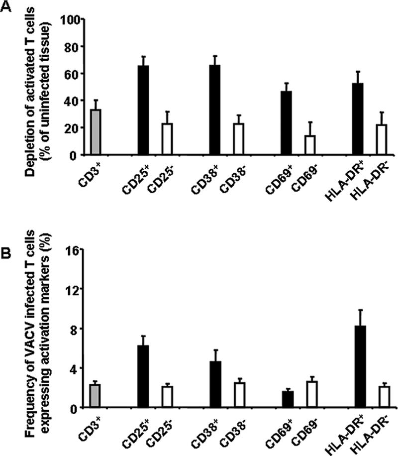
FIG. 3.

Preferential depletion of activated T cells in human lymphoid tissues inoculated ex vivo with VACV. Tissue blocks (27 from each individual human tonsil donor) were infected with NYCBH. At day 12 postinfection, cells were stained for the following activation markers: CD3, CD25, CD38, CD69, and HLA-DR. Data are means ± SEM (error bars) of the numbers of lymphocytes positive or negative for any of the activation markers listed above normalized by the weight of the tissue blocks and by the numbers of corresponding cells in matched uninfected control tissues. A. Depletion of activated CD3+ cells by VACV in human lymphoid tissue ex vivo (n = 12). B. Fraction of VACV-infected activated T cells detected at day 12 postinfection (n = 7).
