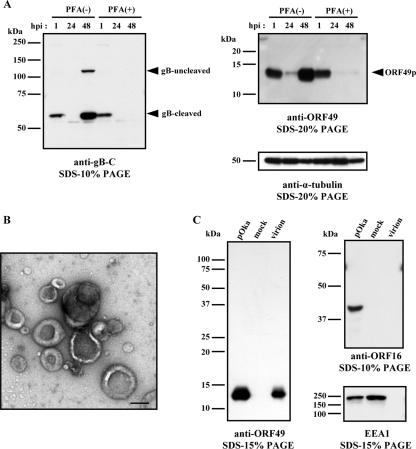
FIG. 1.
Kinetics of protein synthesis and identification of ORF49p in viral particles. (A) MRC-5 cells were infected with pOka cell-free virus at an MOI of 0.1, cultured without (−) or with (+) PFA, and harvested at the indicated time points. The cell lysates were analyzed by WB using Abs to gB and ORF49. An anti-α-tubulin MAb was used as the control. The arrowheads indicate the positions of each protein. (B and C) MRC-5 cells were infected with pOka cell-free virus and cultured until CPE was observed. Histogenz gradient-purified virions were negatively stained with 3% uranyl acetate for electron microscopy (bar, 200 nm) (B) and subjected to WB analysis using Abs to ORF49, ORF16, or EEA1 (C). Molecular mass markers are shown on the left of each panel (A and C).

