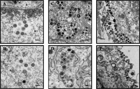
FIG. 6.
Ultrastructural analysis of wild-type and mutant viruses. MRC-5 cells were infected with rpOka (A, C, and E) and rpOkaΔ49 (B, D, and F) by cell-cell spreading and harvested when CPE was observed in almost all cells. The harvested cells were fixed and processed for electron microscopy. Nucleocapsids were observed in the nucleus (n) (A and B), and viral particles were in the cytoplasm (c) (C and D) and on the cell surface (E and F). Bars, 200 nm.

