
FIG. 8.
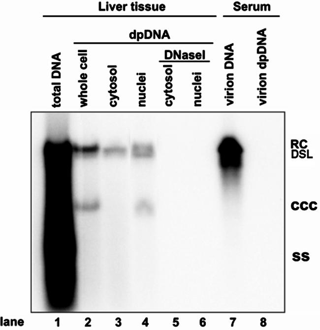
Subcellular distribution of DHBV DP-rcDNA in the liver of virally infected duck. Total intracellular capsid DNA (lane 1), Hirt DNA extracted from total lysate (lane 2), or cytoplasmic and nuclear fractions of a DHBV-positive duck liver without (lanes 3 and 4) or with (lanes 5 and 6) prior DNase I digestion and DHBV DNA extracted from 250 μl of DHBV-positive duck serum with (lane 7) or without (lane 8) prior pronase digestion were resolved in a 1.5% agarose gel, transferred onto membrane, and hybridized with a DHBV minus-strand DNA-specific riboprobe. Total DNA loaded in lane 1 was prepared from 7 mg of duck liver tissue, and each Hirt DNA sample was prepared from lysate prepared from 15 mg of liver tissue. RC, rcDNA; CCC, cccDNA; SS, single-strand DNA; DSL, double-strand linear DNA.