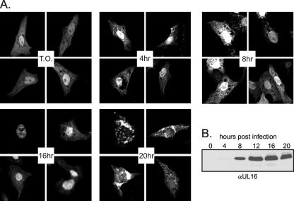
FIG. 1.
Change in UL16-GFP localization during the course of infection. (A) Vero cells were transfected with pUL16-GFP. At 20 to 22 h posttransfection, cells were infected with HSV, fixed with paraformaldehyde at the indicated times postinfection, and viewed by confocal microscopy. T.O., transfected only. (B) Vero cells were infected with HSV, and at the indicated times postinfection, they were harvested and resuspended in sample buffer. Proteins were separated by SDS-PAGE in 10% gels, transferred to nitrocellulose, and detected by immunoblotting with a polyclonal antibody raised against UL16.

