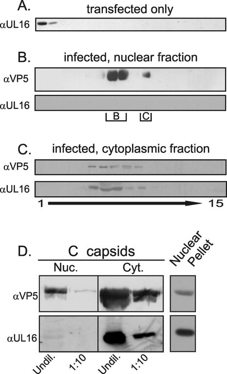
FIG. 2.
Cosedimentation of UL16 with cytoplasmic but not nuclear capsids. Vero cells were transfected with pUL16 (A) or infected with HSV at an MOI of 10 (B to D) for 16 to 22 h (representative 18-h time point shown). Detergent lysates of the transfected cells were analyzed directly in a 20 to 50% (wt/vol) sucrose gradient whereas nuclear and cytoplasmic capsids from the infected cells were first pelleted through a 30% sucrose cushion prior to sedimentation. Fractions were collected, and proteins were concentrated by TCA precipitation. The precipitates were dissolved in sample buffer and separated either in a 7% (for detection of VP5) or 10% (for UL16) polyacrylamide-SDS gel prior to immunoblot analyses with anti-VP5 or anti-UL16 rabbit polyclonal antibodies. The locations of B and C capsids in the gradients are indicated. (D) To compare the levels of UL16 present on equal amounts of nuclear (Nuc) and cytoplasmic (Cyt) C capsids, the relevant species were collected from the gradients, repelleted through a 30% sucrose cushion, and resuspended in sample buffer. Undiluted (Undil) or 1:10 dilutions of the samples were analyzed by immunoblotting with anti-VP5 and anti-UL16 sera. α, anti.

