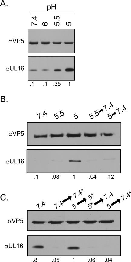
FIG. 5.
Effects of low pH on the association of UL16 with capsids. Extracellular virions were harvested 20 to 24 h postinfection, pelleted through a 30% sucrose cushion, and resuspended in medium buffered to the indicated pH values. (A) After an incubation of 1 h at 37°C, the viral envelopes were removed with NP-40 treatment for 10 min at room temperature. The released capsids were pelleted, dissolved in sample buffer, and analyzed by immunoblotting with anti-VP5 and anti-UL16 antibodies. (B) Virions were first incubated at the indicated pH values for 30 min at 37°C and then incubated for an additional 30 min, either at the same pH or at pH 7.4 (following titration with NaOH). The viral envelopes were removed with NP-40, and the released capsids were collected and analyzed by immunoblotting. (C) Virions were treated as described for the previous panel except that the timing of the NP-40 treatment was either before or after neutralization with NaOH, as indicated by the asterisks. Relative levels of UL16 quantitated by densitometry and normalized for VP5 are indicated bellow each figure. α, anti.

