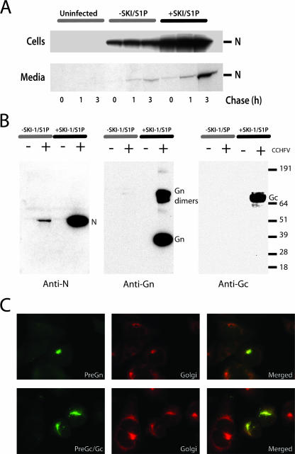
FIG. 4.
Glycoprotein-deficient viral particles are released in the infected cell supernatant of SKI-1/S1P-deficient cells. SRD12B cells stably expressing SKI-1/S1P were produced by transfection of the pIR-SKI-1/S1P vector with Lipofectamine 2000. Two days after transfection, cells were placed in Dulbecco's modified Eagle's medium-F-12 medium supplemented with 5% lipoprotein-deficient fetal bovine serum and penicillin-streptomycin for 14 days to select cells stably expressing SKI-1/S1P. The cell pools growing in the absence of exogenous cholesterol were used in all experiments. (A) SKI-1/S1P-deficient cells (−SKI/S1P) or SKI-1/S1P-deficient cells stably expressing SKI-1/S1P (+SKI-1/S1P) were infected with a multiplicity of infection (MOI) of 5. Twenty-four hpi, cells were pulsed with 100 μCi/ml of [35S]Cys for 30 min and chased for the indicated number of hours, as described previously (22). Triton X-100 was added to a concentration of 1% to cell supernatants prior to the immunoprecipitation with anti-N (5G2) MAb and protein A Sepharose (Amersham). Cell monolayers were solubilized with radioimmunoprecipitation assay buffer and immunoprecipitations carried out as described previously (22). All immunoprecipitated proteins were resolved with a NuPAGE gel system (3 to 8% bis-Tris gels) under reducing conditions. Protein bands were detected using phosphor screens and image digitized and quantitated using a Storm imager (GE Healthcare Biosciences) and ImageQuant software (GE Healthcare Biosciences). At the time of the pulse, we estimated by IFA that ∼15% of the SKI-1/S1P-deficient cells were infected, compared to ∼40% for cells expressing SKI-1/S1P (data not shown). (B) Confluent 175-cm2 flasks of SRD12B cells or SRD12B cells stably expressing SKI-1 were infected with CCHFV with an MOI of 5. Uninfected (−) and infected (+) cell supernatants were changed 24 h after the infection and collected at 48 hpi and viral particles purified as essentially as described previously (1). Purified viral pellets were solubilized in 100 μl of 2× NuPAGE sample buffer and gamma irradiated with 2 × 10⁶ rads prior to electrophoresis. A portion (20 μl) of the reduced samples was analyzed by Western blotting with chemiluminescence to detect CCHFV structural proteins N (9D5 MAb), Gn (Gn ectodomain polyclonal antibody), and Gc (11E7 MAb). The values at right are markers in kilodaltons. (C) SKI-1/S1P-deficient cells were infected and processed by IFA with antibodies 7F5 (PreGn/GP38) or 11E7 (PreGc/Gc) and Giantin (Golgi) from Covance (Berkeley, CA) 7 hpi. The green signal (anti-mouse Alexa 488) is specific for PreGn (top) or PreGc/Gc (bottom) and red (anti-rabbit Alexa 594) for Giantin (Golgi). Red and green digital images were overlaid (merged) to emphasize the colocalization of PreGn and PreGc/Gc with the cis and median Golgi cisternae marker. Images were acquired with a 100× objective.

