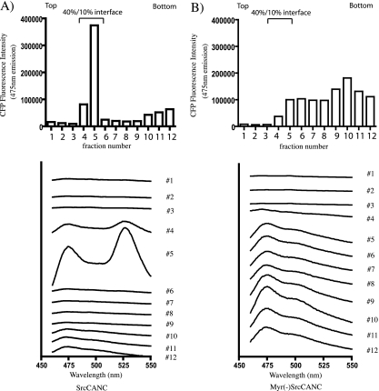
FIG. 3.
Membrane flotation centrifugation of SrcCANC (A) and Myr(−)SrcCANC (B). Cell lysates were layered on the bottom of a 50%-40%-10% iodixanol gradient, and equilibrium flotation centrifugation was carried out to separate membranes (40%-10% interface) from cytosol (fractions 6 to 12). Top bars indicate CFP fluorescence as a marker of total Gag protein in the membrane or cytosol. Bottom curves represent emission scans for each individual gradient fraction.

