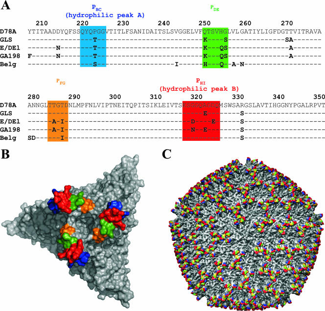
FIG. 3.
Comparison of the amino acid sequences of the variable region of IBDV segment A. (A) Amino acid sequences (aa 206 to 350) of IBDV strains D78, GLS, and E/Del, the Belgian isolate, and isolate GA198 (aa 216 to 350) were compared. The sequences of D78, GLS, and E/Del were taken from the report of Vakharia et al. (36). The sequence of isolate GA198 was taken from GenBank (accession no. AAV48814). The sequence of the Belgian isolate was established in this study. The two hydrophilic peaks A and B, as described previously (1), are shown (see the text). Loops PBC, PDE, PFG, and PHI of domain P of VP2 (6) are boxed in the sequence alignment in blue, green, yellow, and red, respectively. Amino acid sequences are numbered in accordance with the work of Mundt and Müller (23). (B) Surface representation of the trimer, with each surface loop of domain P colored according to the alignment presented in panel A. (C) Locations of the trimers and antigenic loops in the overall structure of the virus particle.

