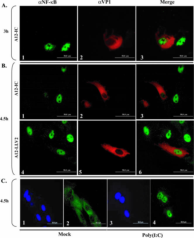
FIG. 3.
p65/RelA indirect immunofluorescence analysis during FMDV infection. PK cells were infected at an MOI of 10 with either WT (A and B, panels 1 to 3) or leaderless (B, panels 4 to 6) virus. Parallel mock infection (C, panels 1 and 2) or poly(I:C) treatment (25 μg/ml and Lipofectamine) (C, panels 3 and 4) was performed. p65/RelA was detected using a rabbit polyclonal antibody (Abcam RB-1638) recognizing the C terminus of p65/RelA and an Alexa Fluor 488-conjugated secondary antibody. The viral protein VP1 was detected using mouse monoclonal anti-VP1 antibody (αVP1) (6HC4) and an Alexa Fluor 594-conjugated secondary antibody. Nuclei were stained with DAPI (C, panels 1 and 3).

