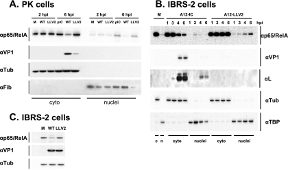
FIG. 6.
WT FMDV infection leads to a loss of p65/RelA. PK (A) or IBRS-2 (B) cells were infected at an MOI of 10 with WT or leaderless FMDV for the indicated times. IBRS-2 (C) cells were infected for 6 h with WT virus (MOI of 10) or leaderless virus (MOI of 100). At the indicated times, cytoplasmic (cyto or c) and nuclear (nuclei or n) extracts (A and B) or whole-cell extracts (C) were prepared and analyzed by Western blotting using rabbit polyclonal-anti p65/RelA antibody (RB-1638), mouse monoclonal anti-α-tubulin antibody (αTub) (Ab-2 MS-581), mouse monoclonal anti-viral VP1 antibody (6HC4), rabbit polyclonal anti-leader antibody, mouse monoclonal anti-fibrillarin antibody (αFib) (ab4566), and mouse monoclonal anti-TBP antibody (ab818). M, molecular weight marker.

