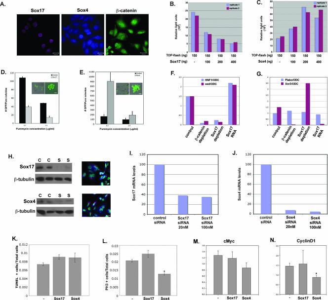
FIG. 2.
Sox17 and Sox4 have opposite effects on Wnt activity and proliferation of SW480 colon carcinoma cells. (A) Endogenous Sox17, Sox4, and β-catenin proteins are present in SW480 cells. (B) Sox17 represses endogenous Wnt signaling activity in colon carcinoma cells. Transfection with the TOP-flash reporter alone (lane 1) demonstrates that SW480 cells have high levels of endogenous Wnt/β-catenin/TCF activity. Cells cotransfected with increasing concentrations of a Sox17 expression plasmid (100 to 400 ng) showed dose-dependent reduction in endogenous Wnt activity (lanes 2 to 4). TOP-flash activity is expressed in relative light units that were normalized to the transfection efficiency by using a renilla plasmid. The results shown are representative of those from at least three independent experiments. −, absent. (C) Sox4 enhances endogenous Wnt signaling activity in colon carcinoma cells. SW480 cells that were cotransfected with increasing levels of a Sox4 expression plasmid exhibited a dose-dependent increase in TOP-flash activity. Results for replicate samples are shown, and experiments were done three times. −, absent. (D) Sox17 represses the proliferation of SW480 cells. The formation of puromycin-resistant SW480 cell colonies was used to measure the effect of Sox17 on cell proliferation. SW480 cells were cotransfected with the pBABE-puromycin resistance plasmid, the pmax-GFP plasmid, and either an empty vector or a Sox17 expression vector and grown in puromycin-containing medium for 5 days. Insets show GFP-positive cells before (left) and after (right) puromycin selection. Only colonies that had ≥10 GFP-positive cells were counted. Error bars indicate standard errors of the means of results for three replicate samples. Puro, puromycin. (E) Sox4 promotes the proliferation of SW480 cells. SW480 cells that expressed Sox4 formed more colonies than vector-alone controls. Insets show GFP-positive cells before (left) and after (right) puromycin selection. (F and G) The effects of the loss and gain of function of Sox17 on Wnt and Sox17 target gene expression in Xenopus embryos. MO (20 ng each) previously used to deplete Xenopus embryos of Sox17 (6) or β-catenin (13) were injected into two-cell embryos, and stage 11 embryos were harvested and pooled in groups of three for each condition. Real-time RT-PCR analysis was used to analyze the relative levels of expression of the Wnt target genes Xnr3 and Siamois (data not shown), the Sox17 target gene Hnf1β, or the ubiquitously expressed gene Plakoglobin (Plako). All gene expression levels are shown as a ratio of the expression of the indicated gene to that of the ubiquitously expressed ODC gene (44). These results demonstrate that Sox17 is required to restrict the activity of Wnt/β-catenin/TCF in Xenopus embryos. (H) siRNA-mediated knockdown of Sox17 and Sox4 proteins in SW480 cells. SW480 cells were transfected with 50 nM scrambled control siRNA (C) or the Sox siRNA (S). The left panels show Western blot analyses of Sox17 and Sox4 proteins, and the right panels are confocal images of transfected SW480 cells showing the typical perinuclear localization of the fluorescently labeled control siRNA (red). Nuclei are stained with SYTOX green. (I and J) siRNA knockdown of endogenous Sox17 and Sox4 mRNA levels. Transfection with Sox17 siRNAs resulted in a 70% reduction of endogenous mRNA, whereas Sox4 siRNAs effected >90% reduction relative to mRNA levels in cells transfected with a control siRNA. (K) Quantitative assessment of apoptosis in SW480 cells as measured by terminal deoxynucleotidyltransferase-mediated dUTP-biotin nick end labeling (TUNEL) staining 48 h after transfection with Sox4 and Sox17 siRNAs. −, control. (L) Quantitative assessment of proliferation of SW480 cells as measured by phosphohistone-H3 (PH3) staining 48 h after transfection with siRNAs. * indicates significance corresponding to a P value of 0.001 versus control samples as calculated using a t test analysis. −, control. (M and N) Quantitative RT-PCR analysis of the Wnt target genes c-myc (I) and cyclin D1 (J) in SW480 cells 48 h after transfection with Sox4 and Sox17 siRNAs. Quantitative PCR analyses of triplicate biological samples were performed. * indicates significance corresponding to a P value of 0.02 versus control samples as calculated using a t test analysis. −, control. Scale bars in panels A, D, E, and H, 20 μm.

