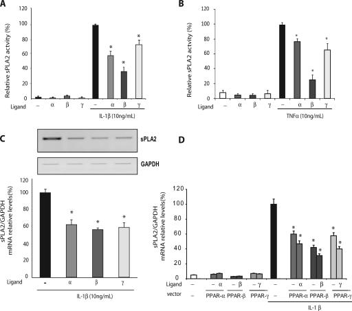
FIG. 1.
IL-1β-induced sPLA2 activity was repressed in response to PPAR ligands. VSMCs were preincubated for 6 h with a specific agonist of PPARα (WY14643; 100 μM), PPARβ (L165041; 50 μM), or PPARγ (GW1929; 1 μM) and then treated or not treated with either IL-1β (A) or TNF-α. (Β) Culture mediums were collected 24 h after IL-1β treatment, and the sPLA2 activity was measured spectrofluorimetrically. (C) Semiquantitative RT-PCR was performed on 1 μg of total RNA from each sample. Amplification was performed with sPLA2 primers and with GAPDH used as a standard gene, as shown in a representative blot. mRNA levels were quantified by the Gene Tools system. (D) VSMCs were transiently transfected with 50 ng of PPARα, PPARβ, or PPARγ expression vector and RXR vector in equimolar quantities. Twenty-four hours after transfection, cells were pretreated for 6 h with a specific agonist of PPARα (WY14643; 100 μM), PPARβ (L165041; 50 μM), or PPARγ (GW1929; 1 μM) and then treated or not treated with IL-1β (10 ng/ml). sPLA2-IIA transcription was measured by reverse transcription/real-time PCR using the expression of the GAPDH gene as an internal standard. Results are representative of three independent experiments. *, P < 0.05 (PPAR agonist-treated versus IL-1β-treated cells). Error bars indicate SEM.

