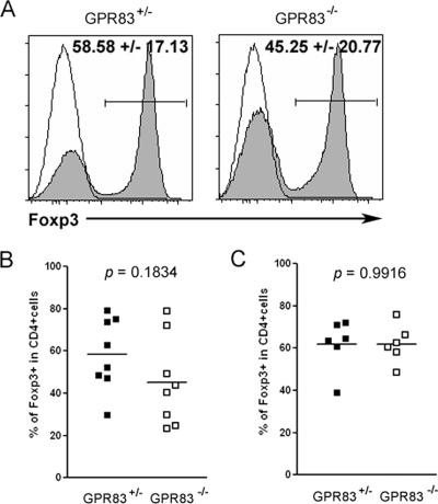
FIG. 4.
Moderate diminution in the efficiency of TGF-β-facilitated Foxp3 induction in the absence of GPR83. (A) Flow cytometric analysis of Foxp3 expression induced in sorted naïve CD25− CD62Lhi CD4+ T cells upon stimulation with anti-CD3 and irradiated splenic APCs in the presence (shaded histogram) or absence (open histogram) of recombinant TGF-β1. Percentages of Foxp3+ cells within the Ly5.1− CD4+-T-cell population are indicated. (B and C) Percentages of Foxp3+ cells induced in the presence of TGF-β1 upon the stimulation of naïve T cells with soluble CD3 antibody in the presence of APCs (B) or plate-bound anti-CD3 and anti-CD28 antibody in the absence of APCs (C). Each symbol represents one independent experiment with two to three mice per group.

