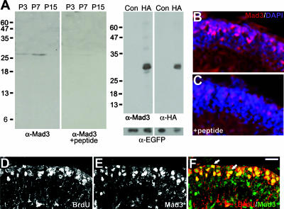
FIG. 2.
Mad3 is expressed in proliferating GNPs. (A) Left panels depict an immunoblot of nuclear extracts prepared from P3, P7, or P15 cerebellum probed with anti-Mad3 antibodies in the absence (left) or presence (right) of Mad3 competing peptide. Right panels depict an immunoblot of HEK293 cell extracts of HA-Mad3- and EGFP-cotransfected (HA) or control EGFP alone-transfected (Con) cells probed with anti-Mad3 (left), anti-HA (right), or anti-EGFP (bottom) antibodies. Molecular mass markers (kDa) are indicated. As expected, HA-Mad3 electrophoreses at a higher molecular mass (29 kDa) than endogenous Mad3 (27 kDa). (B and C) Immunohistochemistry of sagittal sections from P6 cerebellum stained with DAPI (blue) and anti-Mad3 antibodies (red) in the absence (B) or presence (C) of Mad3 competing peptide. Note that the image shown in panel C was exposed 25% longer than the image shown in panel B. (D to F) Immunohistochemistry of sagittal sections of cerebellum from P7 mice injected with BrdU for 2 h and stained with anti-BrdU (D) and anti-Mad3 (E) antibodies. The majority of BrdU+ cells (red) in the EGLa express Mad3 (green) as indicated by arrows in the overlay image (F). Bars, 40 μm (B and C) and 20 μm (D to F).

