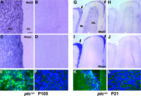
FIG. 6.
Mad3 is upregulated in medulloblastoma and pretumor cells. (A to D and G to J) Sagittal sections of cerebella from 15-week-old ptc+/− mice (P105) that developed tumors (A to D) or from ptc+/− mice at P21 before tumors formed (G to J) were hybridized in situ with DIG-labeled Mad3 (A and G) or Nmyc (C and I) antisense riboprobes. Note that Mad3 and Nmyc transcripts are abundant in ectopic preneoplastic cells (arrows in panels G and I). Sense controls of adjacent sections are shown at right (B, D, H, and J). (E, F, K, and L) Immunohistochemistry of sagittal sections from 15-week-old ptc+/− mice (P105) that developed tumors (E and F) or from ptc+/− mice at P21 before tumors formed (K and L) stained with DAPI (blue) and anti-Mad3 antibodies (green). Mad3 protein is abundant in the tumor tissue (E) and ectopic preneoplastic cells (K); however, staining is absent in normal cerebellar tissue from the same animal (F and L). The area shown in panels F and L is the internal granule layer. Abbreviations: Cb, cerebellum; Mb, medulloblastoma; IGL, internal granule layer; ML, molecular layer. Bars, 200 μm (A to D), 115 μm (G to J), and 30 μm (E, F, K, and L).

