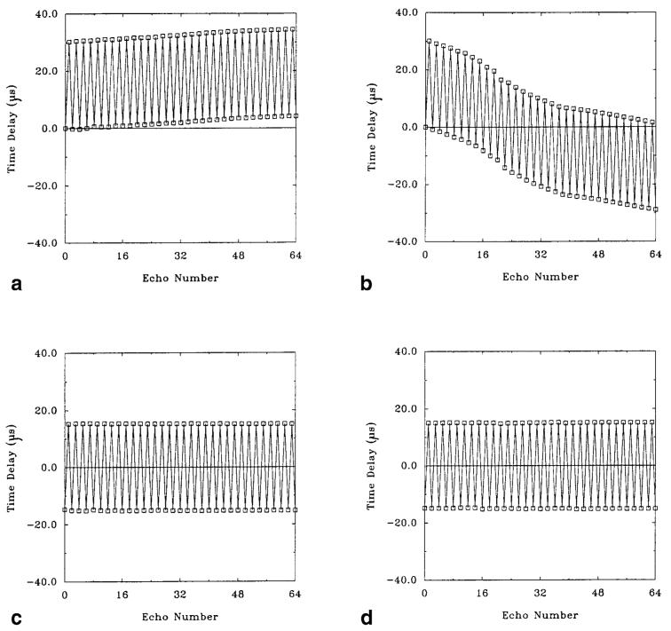
Figure 4.
Time delays (in μsec) plotted against phase-encoding index for a single-shot EPI data set (first 64 echoes). a: Standard reference scan in a water phantom. Time delays were measured with respect to the first echo. b: Standard reference scan in the brain. The trend of the delays is nonlinear, and an average delay is not apparent. c: Balanced reference scan in a water phantom. Time delays were calculated using opposite polarity readout gradients, eliminating the delay bias. The average delay is 15.0 μsec. d: Balanced reference scan in the brain. The data are nearly identical to those obtained in the water phantom, and the average delay is also 15.0 μsec.

