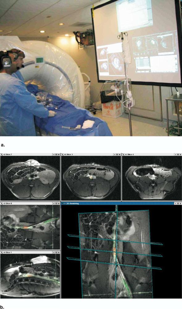
Figure 3.

(a) Picture during an interventional procedure at the National Institutes of Health (NIH). The rear projection screen shows monitors from the MR scanner, an external computer, and the hemodynamics monitor. (b) Real-time multiple slice imaging with active invasive devices and three-dimensional rendering on the custom reconstruction computer at the NIH. Procedure shown is experimental placement of an endograft in an abdominal aortic aneurysm in a pig. Image data from coils in the guiding catheter are highlighted green; image data from coils just distal to the endograft are highlighted red.
